Home/Wellness Zone/Sakra Blogs

9th Jan, 2019

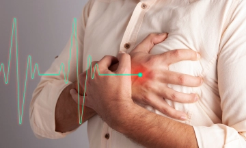
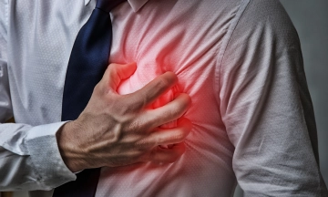
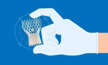
Coronary angioplasty has evolved over the last three decades due to improvements in angioplasty hardware such as stents, balloon catheters, wires etc which have made the procedure far easier to perform. Immediate and long-term outcomes and safety of the procedure have markedly improved. Also aiding in ease of performance are advancements in imaging technologies which have made cardiac Cath labs more sophisticated with lower risk of radiation to both patient and operator. There still are limitations in the two-dimensional image that the angiogram generates which make some interpretations inadequate. We do not get enough detail of what’s happening in the blood vessel wall, and the measurements are not precise enough.
This is where Intravascular Ultrasound comes as a boon. Just like how ultrasound imaging of the intra-abdominal organs can be done using an ultrasound probe, the coronary artery can be imaged in detail using a miniature ultrasound catheter which is taken into the coronary artery during an angioplasty.The image generated by this probe gives multiple details such as the size of the blood vessel, length of the segment to be stented, presence of clot or calcium and such other details which are important in planning the procedure. Also, at the end of the procedure,the IVUS tells us if there are any areas of concern that need to be fine-tuned (such as stent under expansion, under-sizing etc.). This gives a sense of objectivity and completeness to the angioplasty procedure which ensures robust long-term outcomes. The IVUS examination is done without too much addition to procedural time and complexity.
Thus, intravascular imaging a very useful adjunct to angioplasty and helps improve long-term results.

Many everyday habits may seem
8 Jun 2026

When someone suddenly notices
19 May 2026

An arrhythmia happens when
19 May 2026

Feeling tired after a long
19 May 2026

Sleep is not just
24 Mar 2026

Heart failure affects more
6 Mar 2026

Heart disease doesn’t usually
6 Mar 2026

Chest pain is one
6 Mar 2026

Heart failure affects
1 Dec 2025

Heart disease doesn’t usually
1 Dec 2025

Chest pain is one
1 Dec 2025

Feeling a tight sensation in
30 Jul 2025

The terms heart rate and
30 Jul 2025

Heart-related conditions are on the
23 Jun 2025

When it comes to protecting
23 Jun 2025

Coronary Artery Disease occurs when
23 Jun 2025

Heart disease doesn’t happen overnight—and
23 Jun 2025

A heart transplant is a
12 May 2025

HF is the inability of
7 Aug 2024
Heart failure is a serious
6 May 2024

Cardiac arrest is a sudden
6 May 2024

In the realm of chronic
6 May 2024
A heart attack, also known
6 May 2024
The human heart is a
15 Mar 2024
In the realm of advanced
12 Mar 2024
A diagnosis of heart failure
12 Mar 2024
The aorta, the largest artery
25 Aug 2023

A healthy heart is essential
23 Aug 2023
Understanding Heart Failure Heart failure is
14 Aug 2023

Beriberi is also known as
28 Apr 2022

Atrial Fibrillation is a condition
9 Jun 2021

Making resolutions every New Year
17 Jun 2020

Smoking: Should I Use Nicotine
13 Jan 2020

Traveling And Driving After A
10 Jan 2020

Metabolic Syndrome A metabolic syndrome is
10 Jan 2020

Coronary angioplasty has evolved over
9 Jan 2019

Aortic valve surgery is one
27 Nov 2018

What is Coronary Angiography? Coronary angiography
10 Dec 2016

What is a Heart
10 Dec 2016

What is Unstable Angina? Unstable angina
10 Dec 2016

What is Angina? Angina is the
10 Dec 2016

Dr. Jagdish from
9 Jul 2016

Trans fat raises
9 Oct 2015

The first hour
6 Oct 2015

Protecting your heart
25 Sep 2015
 Angioplasty - Sakra World Hospital")
Are
24 Feb 2015

What is Cardiac LIS (Least
14 Jan 2015

We’ve
15 Sep 2014

The Center of
8 Jul 2014

Metabolic Syndrome A metabolic syndrome is
1 Jun 2016
Enquire Now

