

Home/Wellness Zone/Sakra Blogs

30th Oct, 2026
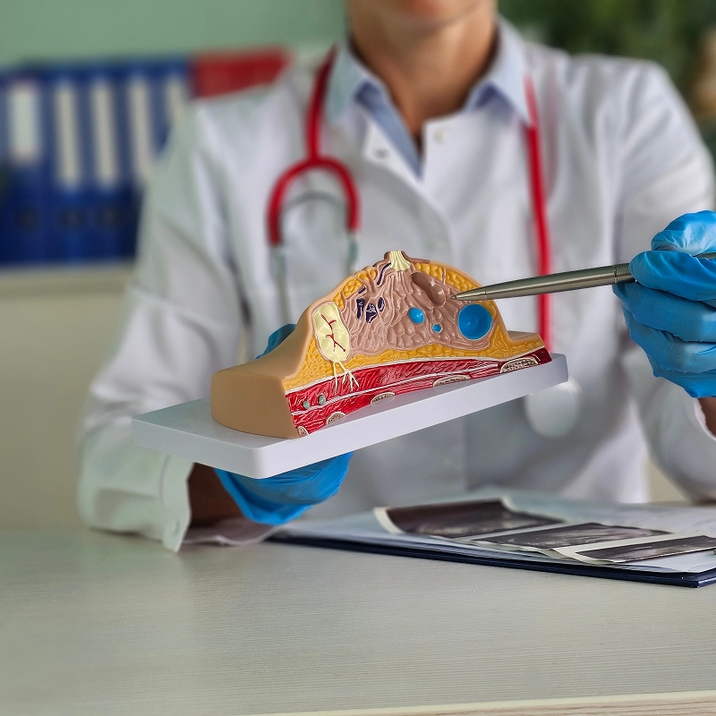
A breast fibroadenoma is a common, non-cancerous lump that develops in the breast. It is made up of glandular and connective tissue and is often found in younger women, particularly those under 30. Fibroadenomas usually feel smooth, firm, and movable under the skin, and they are often painless. Some lumps remain the same size, while others may grow or shrink over time. Although most fibroadenomas are harmless, a new breast lump should always be examined by a healthcare professional to confirm the diagnosis and rule out other conditions. Treatment may involve regular monitoring or surgical removal, depending on the lump’s size, growth, symptoms, and test results.
A breast fibroadenoma is a solid lump formed from the glandular and connective tissues of the breast. It is one of the most common benign breast conditions and is not breast cancer. Fibroadenomas may develop in one or both breasts, and a person can have more than one lump.
These lumps are often round or oval, have clearly defined edges, and move slightly when touched. They may be discovered during a self-examination, a clinical breast examination, or a breast scan.
Fibroadenomas can change with hormonal fluctuations, including those associated with the menstrual cycle, pregnancy, and breastfeeding. Some remain unchanged for years, while others gradually become smaller or disappear.
Breast fibroadenomas do not always cause noticeable symptoms. Many are painless and are first noticed as a lump during bathing, dressing, or a breast examination.
Common symptoms and features include:
A firm breast lump: The lump usually feels round or oval, with smooth, clearly defined edges.
A movable lump: A fibroadenoma often moves slightly beneath the skin when gently pressed.
Little or no pain: Most fibroadenomas are painless, although some people may experience tenderness.
Changes in lump size: The lump may grow, remain stable, or become smaller over time.
Breast tenderness: Some people notice increased tenderness around their menstrual period.
A larger or noticeable lump: A growing fibroadenoma may change the shape or appearance of the breast.
A fibroadenoma usually does not cause skin dimpling, nipple inversion, or bloody nipple discharge. If these changes occur, they should be assessed by a doctor rather than assumed to be related to the lump.
The exact cause of breast fibroadenomas is not fully understood. However, hormones, particularly oestrogen, are believed to influence their development and growth.
Factors associated with fibroadenomas include:
Hormonal changes: Changes in hormone levels during the reproductive years may influence breast tissue growth.
Age: Fibroadenomas are more common in teenagers and younger women, although they can occur at other ages.
Pregnancy: Hormonal changes during pregnancy may cause an existing fibroadenoma to grow.
Hormonal medication: Certain hormone-related treatments may influence the size of a fibroadenoma in some people.
Having a fibroadenoma does not mean a person has breast cancer. Most fibroadenomas are benign, but a proper assessment is needed to confirm the nature of any new breast lump.
Fibroadenomas are classified according to their appearance and the features seen under a microscope. Identifying the type can help doctors decide whether monitoring or removal is appropriate.
A doctor will assess the breast lump through a physical examination and recommend tests based on the person's age, symptoms, and examination findings. The purpose is to confirm whether the lump is a fibroadenoma and distinguish it from other breast conditions.
Common diagnostic tests include:
Clinical breast examination: The doctor checks the lump's size, shape, firmness, movement, and location.
Breast ultrasound: Ultrasound uses sound waves to examine the lump and helps distinguish a solid mass from a fluid-filled cyst. It is often the first imaging test for younger women.
Mammogram: A mammogram uses low-dose X-rays to examine breast tissue and may be recommended depending on age and clinical findings.
Core needle biopsy: A small sample of tissue is collected using a needle and examined in a laboratory when imaging or examination findings need further clarification.
Not every fibroadenoma requires a biopsy. The doctor will decide whether additional testing is needed based on the findings. If the examination, imaging, and biopsy results do not agree, further assessment may be necessary.
Treatment depends on the lump's size, growth, symptoms, and diagnostic results. Many confirmed fibroadenomas do not require surgery and can be monitored over time.
1. Regular Monitoring
Small fibroadenomas that have been confidently diagnosed and are not causing problems may be monitored rather than removed. The doctor may recommend follow-up examinations or imaging to check whether the lump changes.
The timing of follow-up depends on the person's age, test results, and local clinical guidance. Any new or noticeable change should be reported rather than waiting for the next appointment.
2. Surgical Removal
Surgical removal may be recommended if the fibroadenoma is large, growing, painful, causing breast distortion, or uncertain in diagnosis. During the procedure, the surgeon removes the lump while aiming to preserve the surrounding healthy breast tissue.
Surgery is also considered when the patient prefers removal after discussing the potential benefits, risks, and cosmetic effects with the surgeon.
3. Vacuum-Assisted Removal
In selected cases, a fibroadenoma may be removed using a vacuum-assisted device guided by imaging. A small incision is made, and a specialised needle removes tissue from the lump.
This technique may be suitable for certain smaller lumps, depending on their location and other clinical factors. It is not appropriate for every fibroadenoma, and the tissue is usually sent for laboratory examination.
4. Cryoablation
Cryoablation is a procedure that uses extreme cold to destroy the fibroadenoma tissue. A thin probe is placed into the lump under imaging guidance.
It may be an option for carefully selected patients where the diagnosis is confirmed and the procedure is available. Suitability depends on the lump's size, location, and the specialist's assessment.
Not every fibroadenoma needs to be removed. A doctor may recommend removal in the following situations:
Rapid growth: The lump increases in size over a short period.
Large size: The lump is large enough to change the shape or appearance of the breast.
Pain or discomfort: The fibroadenoma causes persistent tenderness or pressure.
Uncertain diagnosis: Imaging or biopsy results do not clearly confirm that the lump is benign.
Suspicion of another condition: The findings raise concern about a different breast lesion, including a phyllodes tumour.
Personal preference: The patient chooses removal after discussing the options with the treating specialist.
The decision should be individualised. Removing a fibroadenoma is not always necessary simply because it is present.
Breast fibroadenoma removal is commonly performed through a procedure called an excisional biopsy or lumpectomy. The exact technique depends on the lump's size, location, and the surgeon's plan.
The procedure generally involves the following steps:
Anaesthesia: Local or general anaesthesia is used depending on the procedure and clinical circumstances.
Small incision: The surgeon makes an incision, often positioned to reduce visible scarring where possible.
Lump removal: The fibroadenoma is separated from the surrounding tissue and removed.
Laboratory examination: The removed tissue is sent for testing to confirm the diagnosis.
Wound closure: The incision is closed with sutures or another suitable method, and a dressing is applied.
Many patients can go home on the same day, although this depends on the procedure and individual circumstances.
Recovery after fibroadenoma removal is usually straightforward, but the time needed varies with the size of the lump and the type of procedure performed. Mild pain, bruising, swelling, or tenderness around the incision may occur for several days.
To support recovery:
Follow the surgeon's instructions for wound care and dressing changes.
Take prescribed pain relief as directed.
Wear a supportive bra if recommended.
Avoid heavy lifting and strenuous upper-body exercise until advised that it is safe.
Attend follow-up appointments to review healing and the laboratory results.
Contact the treating team if pain or swelling increases, the wound becomes red or hot, pus develops, or fever occurs. Heavy bleeding or rapidly worsening symptoms require urgent medical attention.
Most breast fibroadenomas are benign and do not turn into breast cancer. A simple fibroadenoma generally does not significantly increase breast cancer risk.
Some complex fibroadenomas or associated breast tissue changes may be linked to a small increase in risk, depending on the findings and the person's medical history. A fibroadenoma can also sometimes be difficult to distinguish from other breast lumps without appropriate testing.
For this reason, a new breast lump should be assessed, and recommended follow-up should not be skipped. A previous diagnosis of fibroadenoma does not mean that every future breast lump is also a fibroadenoma.
Yes, some fibroadenomas become smaller over time and may disappear without treatment. Others remain the same size for years, while some grow, particularly during periods of hormonal change.
Because the pattern varies from person to person, a lump should not be assumed to be a fibroadenoma without proper assessment. Once the diagnosis is confirmed, the doctor can advise whether monitoring is sufficient or removal should be considered.
Consult a doctor if you discover a new breast lump, even if it is painless or moves easily under the skin. A clinical examination and appropriate imaging can help identify the cause.
Seek medical assessment promptly if you notice:
A lump that grows or changes in shape.
New skin dimpling, redness, or thickening.
A nipple that becomes newly inverted.
Bloody or unexplained nipple discharge.
A lump in the armpit or a noticeable change in breast shape.
These symptoms do not always indicate cancer, but they need evaluation to identify the underlying cause.
A breast fibroadenoma is a common, non-cancerous breast lump that often affects younger women. It may feel firm, smooth, and movable, and it usually does not cause pain. Many fibroadenomas can be safely monitored after the diagnosis is confirmed, while larger, growing, painful, or uncertain lumps may need removal. Clinical examination, breast imaging, and sometimes a biopsy help guide treatment. Anyone who notices a new breast lump or a change in an existing lump should seek medical assessment rather than trying to diagnose it at home.
1. Is a breast fibroadenoma dangerous?
Most breast fibroadenomas are benign and do not pose a serious health risk. However, a new or changing breast lump should be assessed to confirm the diagnosis and rule out other conditions.
2. Can a fibroadenoma be removed without surgery?
Some fibroadenomas can be removed using vacuum-assisted techniques or treated with cryoablation in selected cases. The best option depends on the lump's size, location, confirmed diagnosis, and available expertise.
3. Does breast fibroadenoma removal leave a scar?
Removal surgery usually involves a small incision, so a scar may remain. Its size and appearance depend on the surgical technique, the lump's location, and how the wound heals.
4. Can a fibroadenoma grow during pregnancy?
Yes, hormonal changes during pregnancy can cause some fibroadenomas to grow. A breast lump that changes during pregnancy should be assessed by a healthcare professional.
5. Can breast fibroadenoma come back after removal?
The same fibroadenoma usually does not grow back if it has been completely removed, but new fibroadenomas can develop in the same or the other breast. Follow-up is important if a new lump or change appears.

Senior Consultant & Lead – General Surgery, Laparoscopic & Robotic Surgery
Book an Appointment
Finding a lump in the breast can be worrying, but not every breast lump is a sign of cancer. Many lumps develop because of hormonal changes, fluid-filled cysts, or the growth of normal breast tissue. These are called benign
30 Oct 2026
Gallstones are a common digestive problem that can cause significant discomfort and complications if left untreated. While some people may not experience symptoms, others may develop severe pain and require medical attention. Understanding gallstone surgery, its types, and recovery
21 Jul 2026
Nipple discharge is a condition where fluid comes out of one or both nipples, either on its own or when pressure is applied. While it can be alarming, especially if unexpected, not all cases are serious. Many people often
7 Jul 2026
Breast pain is one of the most common concerns women experience at some point in their lives. It can feel mild, sharp, heavy, or even constant, which often leads to worry. Many people frequently ask questions like why breast
7 Jul 2026
The thyroid is a small, butterfly-shaped gland located at the front of the neck. It plays an important role in controlling metabolism, heart rate, energy levels, and body temperature. When thyroid problems cannot be managed with medicines alone, doctors
12 Jan 2026
A hernia happens when an internal organ or tissue pushes through a weak spot in the muscle or surrounding tissue. It often appears as a visible bulge and may cause discomfort, especially while lifting, coughing, or standing for long
12 Jan 2026
Enquire Now


