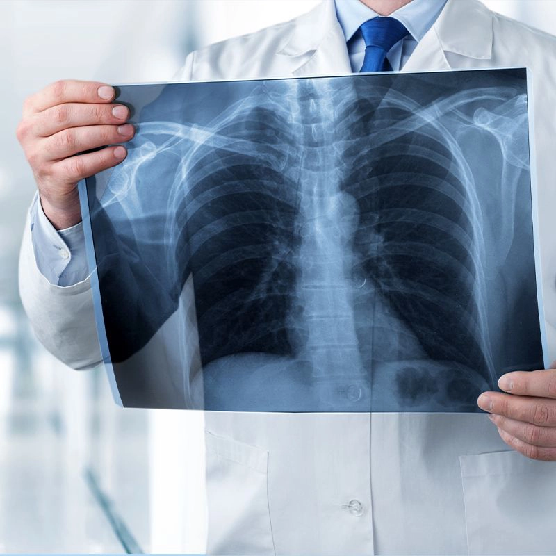
What is Radiology?
Radiology, also called diagnostic imaging, allows the doctor to see inside the body through a series of several tests that take images of various parts of the body. The doctor who is capable of interpreting the images of the body using various different machines is a Radiologist, who is a specialist in this field.
The several imaging exams that are used to provide the image are X-ray, MRI, CT scan, PET scan, Mammogram, Ultrasound, Fluoroscopy, Bone scan, Thyroid scan, thallium cardiac stress test, etc. Sakra World Hospital is one of the best radiology hospital in Bangalore. The department of Radiology & Imaging at Sakra World Hospital offers a complete range of diagnostic and interventional radiology services while maintaining the values of safety and patient-focused care.
What is the use of Radiology?
The different radiology procedures help the doctors to:
-
Diagnose the causes of the symptoms and come to a conclusion as to what disease you may have.
-
Diagnose for different conditions such as breast cancer, colon cancer, liver disease, heart disease, lung diseases such as infection and cancer, etc.
-
Monitor how well your body is responding to a treatment you are receiving for your condition.
What is the difference between a CT scan and MRI imaging procedures?
Just like an X-ray machine, a CT scan machine also uses X-rays to produce images of your internal body parts but provides much more minute detail as compared to X-rays.
CT scan uses X-rays to produce images of the internal structures of the body, while MRI uses powerful magnetic fields and radio waves to produce images of internal body structures.
What is a CT scan and how long does it take?
A CT (Computerized Tomography) scan uses computers and rotating X-ray machines to create images of the body. These images give more detailed information than a normal X-ray. CT scan can show the organs of the body such as the brain, liver, kidneys, soft tissues, blood vessels, and bones in various parts of the body such as the head, shoulders, spine, heart, abdomen, knee, and chest. The whole procedure takes 5 to 20 minutes to complete.
What are the risks associated with CT scans?
The risks associated with CT scan are:
-
Cancer, due to exposure to radiation, but is very rare.
-
Allergic reaction to the contrast material.
-
Multiple CT scans increase the risk of cancer
Why is a CT scan performed?
The CT scan is performed to diagnose diseases and examining injuries. It helps the doctor to:
-
Assess the liver, pancreas, adrenal glands, kidneys, intestines, lungs, brain, heart, cancers of head and neck, etc.
-
Diagnose infections, bone fractures, muscles disorders.
-
Study the blood vessels and other internal structures.
-
Guide procedures such as surgeries and biopsies.
-
Examine the extent of internal injuries and internal bleeding.
-
Monitor the effectiveness of treatments for certain medical conditions like cancer and heart disease.
What happens when you have an MRI?
The MRI machine uses a strong magnetic field around you and radio waves are directed at your body. In some cases, a contrast material is injected into the veins to enhance the images. You must stay still as any movement can blur the images. The MRI lasts from 15 minutes to an hour. MRI provided different information as compared to CT scan and shows the body organs and soft tissue structures in detail.
Sometimes you may need a combination of CT and MRI scans since the information provided by both is different. The doctor and radiologist will tell you whether you need both the tests or not.
What is a Mammogram?
A mammogram is an X-ray of the breast which the doctor uses to look for early signs of breast cancer. Mammograms help doctors to find breast cancer early sometimes even before it can be felt.
What happens if my mammogram is abnormal?
An abnormal mammogram doesn’t always mean that there is cancer. The cancer specialist will order additional mammograms and other tests to confirm the diagnosis.
Why an ultrasound is performed?
Ultrasound uses sound waves to produce images of the body and it has different uses as compared to CT and MRI. Since sound waves do not have as much depth assessment as CT and MRI, an ultrasound can only show some of the information and not all.
An ultrasound is performed if you have any condition that requires an internal view of the organs. An ultrasound can give the image of gall bladder stones, kidney stones (if large), brain (in infants), eyes, liver, uterus, ovaries, pancreas, spleen, thyroid, testicles, and blood vessels.
An ultrasound also guides the Radiologist during certain medical procedures such as biopsies.
Finally, at the end of each type of Radiological test, your images will need to be interpreted in detail by the Radiologist doctor, who will provide the diagnosis and discuss your findings with your primary care doctor.
What you can do to help in your own care and diagnosis is to always carry your medical file with you with all your old test results, old X-rays and CT scans if you have them; and share them with your Radiologist.
At Sakra World Hospital, we have the best MRI scan centre in Bangalore. Backed with the latest equipment and a team of the best radiologists in Bangalore, we offer all types of imaging services.


