To know more log onto www.sakraworldhospital.com
 + 91 91080 13673
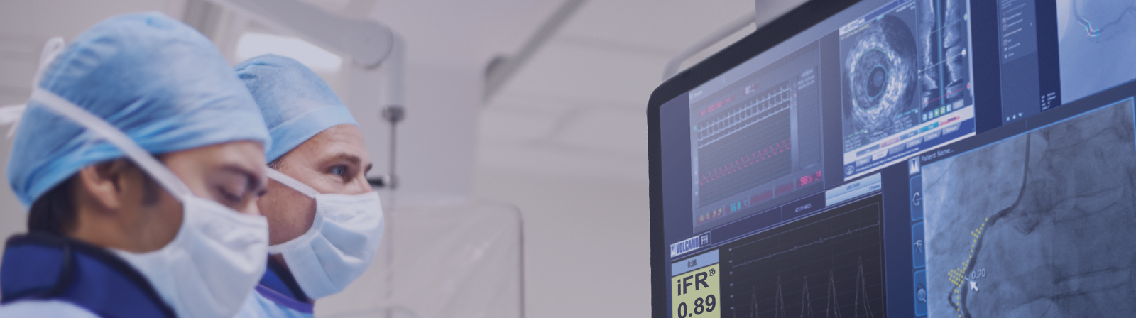
Intravascular Ultrasound (IVUS) allows the doctor to see a coronary artery inside out, which provides real time information that goes beyond routine imaging methods such as coronary angiography or CT scans.
The information provided by IVUS provides for a more accurate placing of the stent, reducing complications and incidence of stent clots.
In IVUS, the miniaturized transducers that is less than four hundredth of an inch, is placed on a catheter. This catheter is slipped along the guide wire used in angioplasty. It becomes a tiny camera that gives the cardiologist a view of the three distinct circular layers of the artery. The cardiologist can make the clinical decision more effectively using this image.


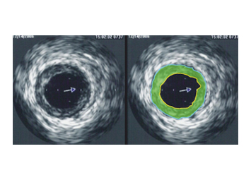
Intravascular ultrasound image of a coronary artery
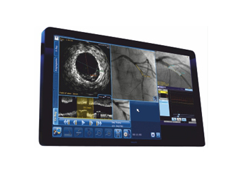
Split screen view of the IVUS monitor
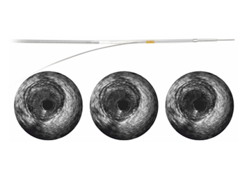
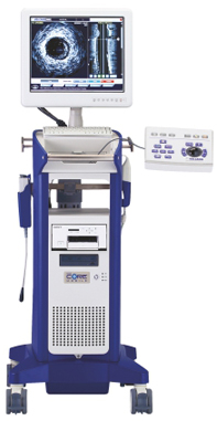
Rotational IVUS catheter & Imaging
Increased accuracy of stent placement
Reduced risk of complications
Quick recovery and improved quality of life
Intravascular Ultrasound (IVUS) allows the doctor to see a coronary artery inside out, which provides real time information that goes beyond routine imaging methods such as coronary angiography or CT scans.The information provided by IVUS provides for a more accurate placing of the stent, reducing complications and incidence of stent clots.
The current "gold standard" of angiography shows only the lumen as an X-ray image created by injection of contrast dye. With IVUS, just a few clicks on the console measures the area of the blockage, the size of the artery and an accurate percentage of narrowing. Vulnerable plaque, which exist inside the arterial wall, also can be visualized by IVUS thereby preventing rupture and potential heart attacks.
For Appointment www.sakraworldhospital.com or Call:080 4969 4969
